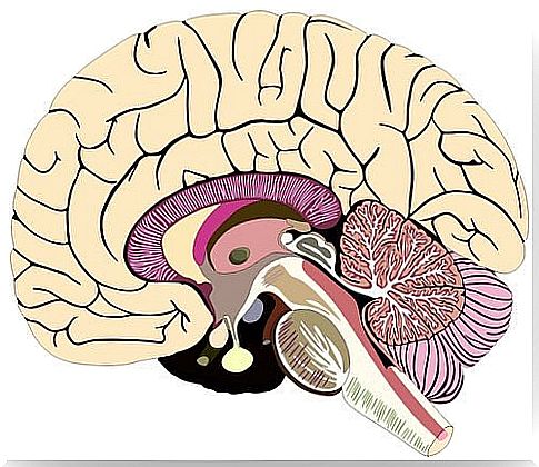
Meninges: Structure And Functions
The brain and spinal cord are surrounded by three membranous layers: the meninges. These are the dura, arachnoid, and pia mater. The combination of the latter two, pia mater and arachnoid, form the leptomeninges, while the dura mater forms the pachymeninges.
The main function of the meninges is to provide a protective layer for the brain, a very vulnerable organ. As a result, it needs special protection that no one else has. At least not in the same way. So the meninges are responsible for this. Furthermore, these protective layers participate in the blood-brain barrier.
The meninges develop from a precursor layer known as the primitive meninges. This is composed of elements derived from the mesenchyme and neural crest. Furthermore, it is separated into two distinct layers: internal endomeninges and external ectomeninges.
The endomeninges differs into arachnoid and pia mater and is derived from both mesoderm and ectoderm. The ectomeninges forms the dura mater and the bones of the neurocranium and is formed only from the mesoderm.
structure of meninges
the dura mater
It is the outermost layer. The dura is composed of two layers. The first, the outer layer, is the periosteum of the skull and contains blood vessels and nerves. Adheres to the inner surface of the skull with specially fitted joints to the sutures and base of the skull.
The deepest layer of the dura is known as the meningeal layer. This layer is responsible for forming reflexes that divide the brain into compartments.
Among these compartments, the most prominent are the sickle brain and the tent cerebellum. Furthermore, there is no distinctive border between the meningeal dura and the periosteum. This only occurs when they separate to form the dural venous sinuses. The layers can be differentiated histologically by the fact that the meningeal layer has fewer fibroblasts and proportionally less collagen (2).
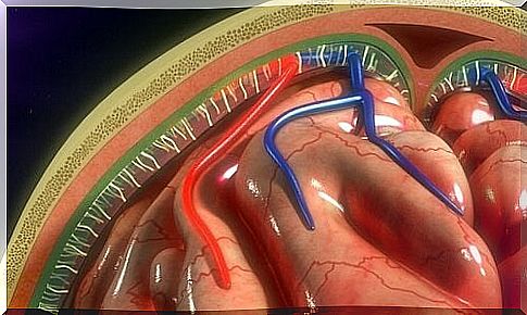
Arachnoid or intermediate layer
The arachnoid is the middle layer of the meninges. Contains the subarachnoid space which, in turn, stores the cerebrospinal fluid (CSF). The depth of the subarachnoid space varies depending on the relationship between the arachnoid and pia mater layers.
This layer is made up of two different cell layers. Following the edge of the dura mater cells is the layer of arachnoid barrier cells (3). This layer is full of cells tightly bound together by countless desmosomes and tight junctions. Thus, these provide the layer with a barrier function that prevents fluid movement through it.
At the bottom of the arachnoid is the reticular arachnoid layer. Cells in this layer join the subarachnoid space and join the pia mater. Furthermore, they involve the blood vessels that pass through the layer (1).
- Arachnoid granulations. They are microscopic structures that play an important role in CSF absorption. However, the mechanism is unclear. Furthermore, it is considered that arachnoid granulations may also play a role as a regulator of CSF volume.
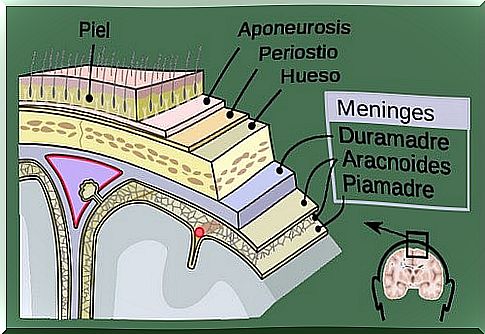
pia mater
The pia mater is the innermost layer of the meninges. It is a delicate, well-vascularized, connective tissue structure that surrounds and protects the brain and spinal cord.
It forms a continuous layer of cells well adhering to the surface of the brain that submerge in fissures and grooves. Cells are connected by desmosomes and gap junctions, which, as a result, allows this layer to fulfill a barrier function.
Virchow-Robin spaces
Virchow-Robin spaces are spaces around the (perivascular) vessels that surround the small arteries and arterioles. They pierce the surface of the brain and extend into the subarachnoid space (1).
These spaces have been shown to increase in size with age, without an associated apparent loss of cognitive function (4). Furthermore, the dilation of these spaces is associated with pathologies such as hypertension, neuropsychiatric disorders, multiple sclerosis and trauma (5).
To conclude, the authors Patel and Kirmi (2009) emphasize the importance of knowing the meninges. Thus, it is essential to understand the structure, functions and anatomy: this will allow us to understand the spread and location of pathologies related to meninges. Furthermore, the most common pathology known in relation to these is meningitis.